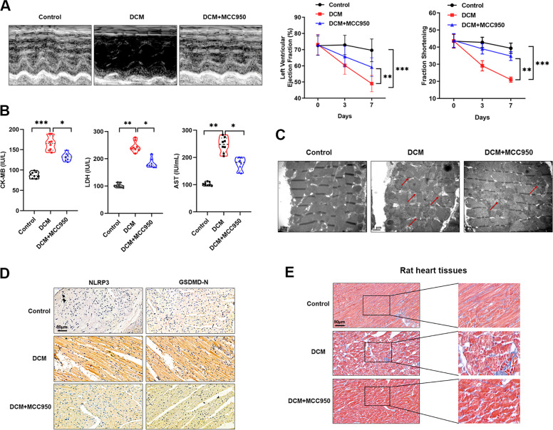
Fig. 2. Pyroptosis was involved in DCM in vivo.
A Morphological changes (left panel) and quantitative analysis of LVEF and FS (right panel) in rat hearts were detected by echocardiography, **P < 0.01, ***P < 0.001. B Serum expression of myocardial enzymes CKMB, LDH and AST were quantified in rats of respective groups, *P < 0.05, **P < 0.01, ***P < 0.001. C A representative section of the left ventricle from respective groups of rats showing sporadic mitochondrial, damaged disconnected myofibers, and thinner myofibers. D IHC analysis was performed to detect the expression of NLRP3 and GSDMD-N in hearts of DCM rat after treated with MCC950. E Masson staining was done in heart tissues of DCM rats treated with MCC950.

