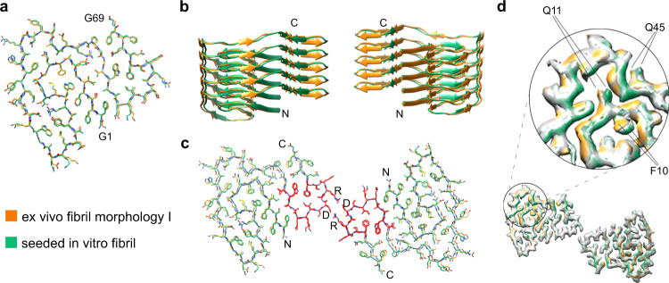
Fig. 4. Comparison of the major seeded in vitro fibril and the ex vivo fibril morphology I.
a Superimposition of the molecular models of the fibril protein in the two fibrils. All atom RMSD: 0.81 Å. b Superimposition of ribbon diagrams of five-layer stacks of the two fibrils. c Superimposition of one molecular layer of the two fibrils. The all atom RMSD values are 1.09 Å for residues 1–69, 0.46 Å for residues 51–64 (red) and 1.17 Å for residues 1–50 and 65–69. In all three panels: light green, major seeded in vitro fibril; orange, ex vivo fibril. d Comparison of the 3D maps of the two fibrils (difference map). The green and orange regions refer to the difference between the two maps.

