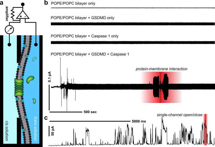
Fig. 2. In vitro characterization of GSDMD pores in lipid bilayers reveals repeated spontaneous pore opening and closing events.
a Schematic for electrophysiological setup used to monitor reconstituted GSDMD pores in a planar lipid bilayer in vitro. Silver-silver chloride electrodes connecting cis and trans compartments conducted the current, which was subsequently amplified and recorded. b Representative traces of GSDMD protein–membrane interaction and single pore formation events in lipid bilayers using a gap-free protocol. From the top of the panel: recordings in controls including baseline bilayer (POPE/POPC) alone, baseline bilayer with the addition of GSDMD protein only, and baseline bilayer with the addition of caspase-1 only, were absent of current activity. Bottom of panel: addition of activated GSDMD to the baseline bilayer demonstrated protein–membrane interaction characterized by fluctuating micro-currents. c Representative trace of single pore activity showing spontaneous and repeated opening and closing events across the GSDMD pore.

