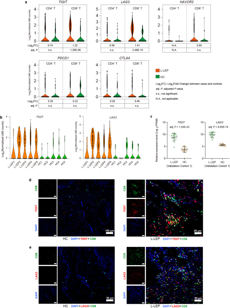
Fig. 4. CD8+ T cells in L-LEP lesions showed an exhausted state in L-LEP patients.
a Expressions of TIGIT, LAG3, HAVCR2, PDCD1, and CTLA4 in skin CD4+ and CD8+ T cells of the discovery cohort. b Expressions of TIGIT and LAG3 in the skin CD8+ T cells of each sample. c Expressions of TIGIT and LAG3 in the skin by cases and controls of validation cohort 1 as determined by bRNA-seq. d Co-localization of TIGIT and CD8 in L-LEP lesions. The nucleus was stained by DAPI. e Co-localization of LAG3 and CD8 in L-LEP lesions. The nucleus was stained by DAPI.

