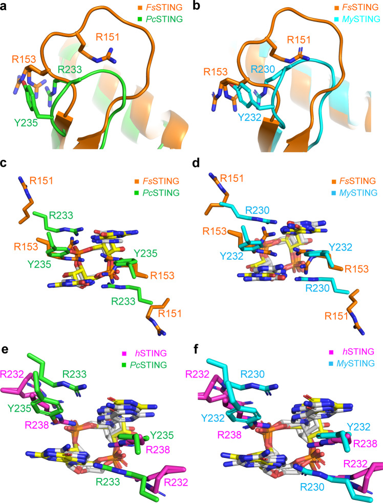
Fig. 3. Structural comparison of the β-strand lids and the ligand-binding pockets of bacterial STING and metazoan STING.
a, b Superimposition of the β-strand lids from the 3’,3’-cGAMP-bound FsSTING (orange, PDB: 6WT4) and (a) PcSTING/c-di-GMP complex (green) or (b) MySTING/c-di-GMP complex (cyan). c, d Enlarged view of the ligand-binding pocket of FsSTING superimposed with (c) PcSTING or (d) MySTING. The specificity-determining residues in β-strand lids are shown as sticks and labeled. C-di-GMP and 3’,3’-cGAMP are shown as yellow and white sticks, respectively. e, f Enlarged views of the ligand-binding pockets following superimposition of the 2’,3’-cGAMP-bound hSTING (magenta, PDB: 4KSY) with (e) PcSTING/c-di-GMP complex (green) or (f) MySTING/c-di-GMP complex (cyan). The specificity-determining residues in PcSTING, MySTING and hSTING are shown as sticks and labeled. C-di-GMP and 2’,3’-cGAMP are shown as yellow and white sticks, respectively.

