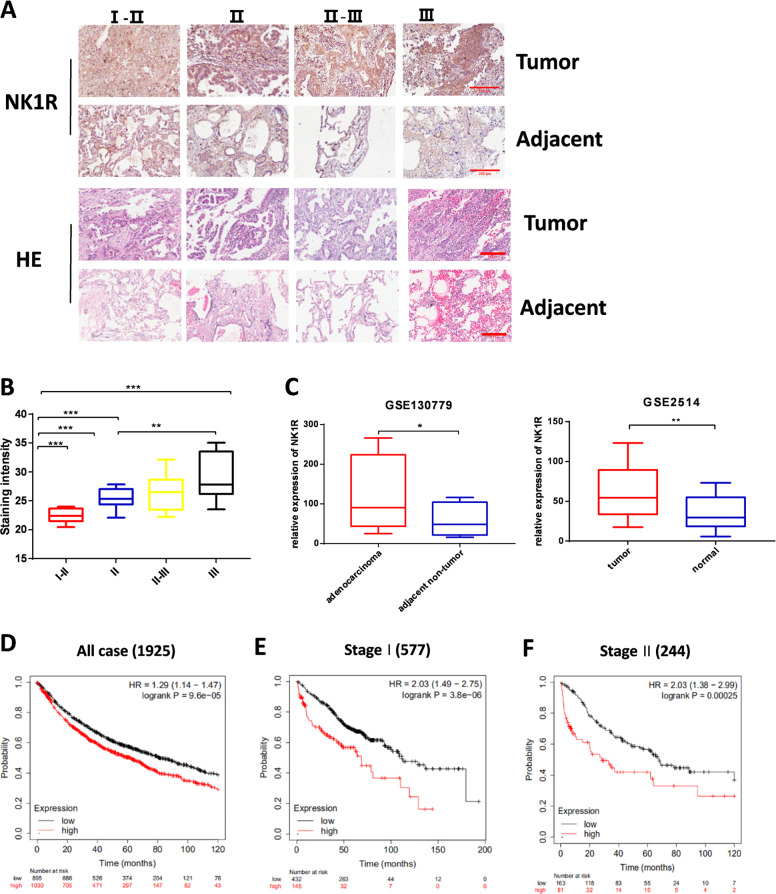
Fig. 1. Expression level of NK1R is related to NSCLC progression and poor prognosis.
IHC staining of NK1R was performed on tissue microarrays of 30 human lung adenocarcinoma patients. A The IHC analysis of NK1R expression in tumor tissues compared with matched adjacent tissues from stage I–II (n = 3), stage II (n = 11), stage II–III (n = 13), to stage III (n = 3). Scale bar, 200 μm. B IHC staining intensity of NK1R in tumor tissues of lung adenocarcinoma from stage I to stage III. C NK1R expression analysis in human lung cancer in comparison with the matched adjacent tissues in GSE130779 and with normal tissues in GSE2514. D–F The Kaplan–Meier overall survival analysis with the log-rank test in 1925 cases from GEO and TCGA datasets (see details in Methods), defined by tumor stages and by low/high NK1R expression. *P < 0.05, **P < 0.01, ***P < 0.001.

