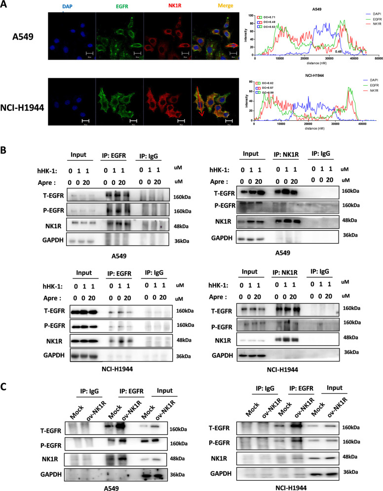
Fig. 6. NK1R interacts with EGFR in NSCLC cells.
A Confocal microscopy analysis to detect the colocalization of EGFR and NK1R in A549 and NCI-H1944. Green, EGFR; Red, NK1R; nucleus was stained by DAPI. Scale bar, 20 μm. The plot profile of fluorescence intensity was used to show the overlap of the NK1R and EGFR. Overlap Coefficient (OC) was >0.6 as the indication colocalization, while overlap coefficient was <0.6 as the absence of colocalization. B Co-immunoprecipitation assay by the antibody of NK1R or EGFR to show NK1R-EGFR interaction in A549 and NCI-H1944 cell lines. Cells were treated with hHK-1 or aprepitant for 24 h to detect the effect on NK1R-EGFR interaction. C Overexpression of NK1R in A549 and NCI-H1944 enhanced NK1R-EGFR interaction in a co-immunoprecipitation assay.

