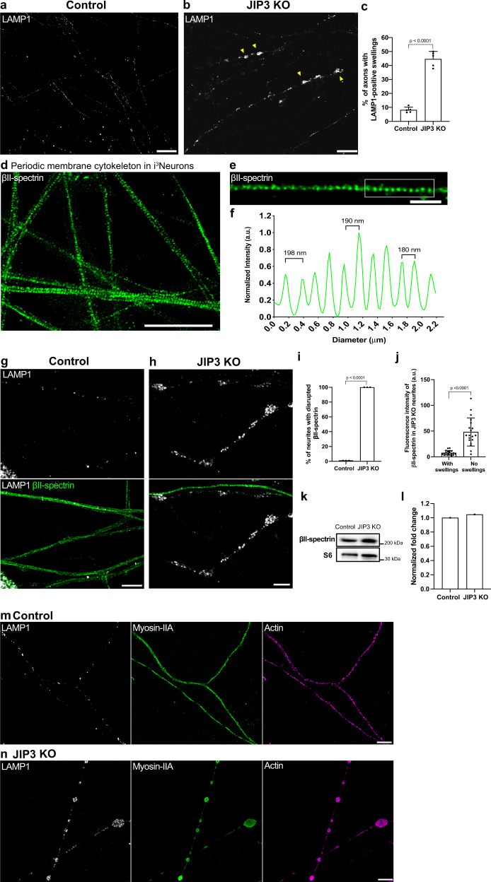
Fig. 1. Lysosome-filled axonal swellings in JIP3 KO axons correlate with global disruption of the membrane periodic skeleton.
a, b Airyscan imaging of control i3Neurons and JIP3 KO i3Neurons (13 days of differentiation). Yellow arrowheads highlight lysosome-positive axonal swellings in the KO neurons (scale bars, 15 μm). c Percentage of i3Neurons containing at least one lysosome-positive axonal swelling represented as mean ± SD, pooled from four independent experiments (n ≥ 32 per experiment, 13 days of differentiation). d, e STED microscopy images of βII-spectrin immunofluorescence in the axons of control i3Neurons (day 17). (Scale bars, d: 5 μm; e: 1 μm). f Graph demonstrating the longitudinal distance between peaks in the βII-spectrin signal from the boxed region in (e). g Airyscan microscopy images of control i3Neurons show regular distribution of lysosomes (LAMP1, white) and intact periodic membrane skeletons (βII-spectrin, green). h JIP3 KO i3Neurons (day 15) exhibit disruption in the spectrin organization in axons positive for lysosome accumulations (scale bars, 5 μm). i Percentage of swollen axons with disrupted periodic membrane skeleton represented as mean ± SD pooled from three independent experiments (≥20 axons analyzed per experiment). Lysosomes and the periodic membrane skeleton were labeled with LAMP1 and βII-spectrin antibodies, respectively. j Graph depicting the mean βII-spectrin fluorescence intensity of JIP3 KO neurites with and without lysosome-filled axonal swellings (≥35 µm in length) represented as mean ± SD, pooled from three independent experiments (n ≥ 20 in total). Note the possible contribution of some dendrites to the “No swellings” group. k, l Immunoblots showing levels of βII-spectrin in control and JIP3 KO i3Neurons (day 15); ribosomal protein S6 was used as loading control (k), and their normalized expression levels is shown in (l). m STED microscopy images of myosin-II filaments (green) show a periodic distribution similar to that of F-actin (magenta) in control i3Neurons. n Myosin-II and actin organization is lost in JIP3 KO i3Neurons (day 15) and both were enriched at the lysosome-positive axonal swellings (white). Lysosomes and myosin-II filaments were labeled with antibodies against LAMP1 and non-muscle myosin-IIA respectively, while rhodamine phalloidin was used to label F-actin. Scale bars, 5 μm. p-values were calculated using two-tailed Student’s t-test.

