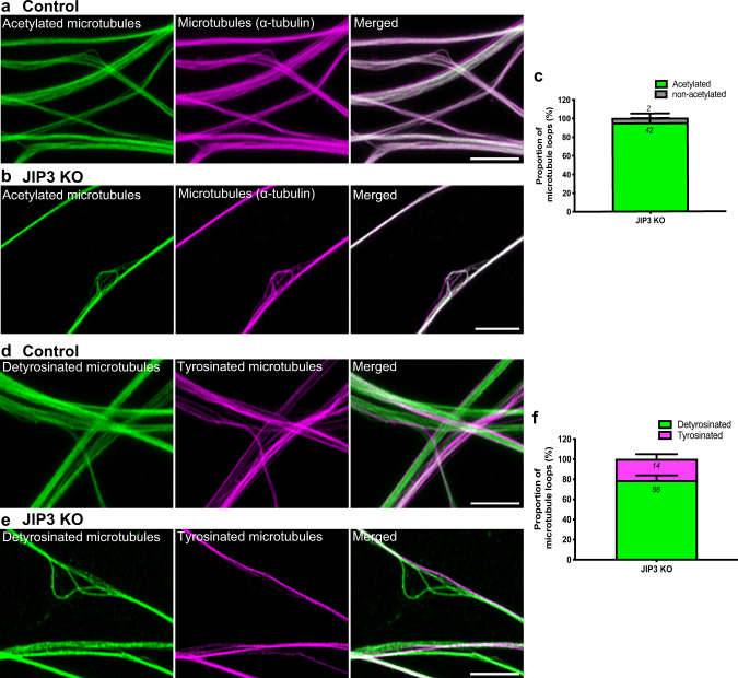
Fig. 3. Microtubule loops in JIP3 KO axonal swellings are primarily detyrosinated.
a, b Airyscan microscopy images of acetylated-α-tubulin, (green) and total microtubules (magenta) in control and JIP3 KO i3Neurons (day 13) respectively. Scale bars, 5 μm. c Fraction of acetylated and non-acetylated microtubules in JIP3 KO i3Neurons (pooled data from 2 experiment with ≥19 swellings analyzed per experiment). d, e Airyscan microscopy images of neurites from both control (d) and JIP3 KO i3Neurons (day 13) (e) consist of parallel microtubule bundles that are either detyrosinated (green) or tyrosinated (magenta) in control and JIP3 KO i3Neurons, respectively. Scale bars, 5 μm. f Fraction of detyrosinated and tyrosinated microtubules in JIP3 KO i3Neurons (pooled from three independent experiments with ≥18 swellings analyzed per experiment).

