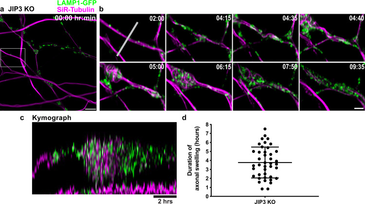
Fig. 4. Lysosome-positive axonal swellings are dynamic in JIP3 KO neurons.
a Representative image of JIP3 KO i3Neurons (day 15) stably expressing LAMP1-GFP (green) and labeled with SiR-tubulin (magenta) at the beginning of a 12-h time lapse imaging time course (Airyscan microscopy). Scale bar, 5 μm. b Images from the boxed area in (a) at the indicated time points. Scale bar, 2 μm. c Kymograph of the region (marked by white line in b; scale bar, 2 h). d Scatter dot plot (mean ± SEM) depicting the duration of 41 swellings pooled from 7 independent experiments.

