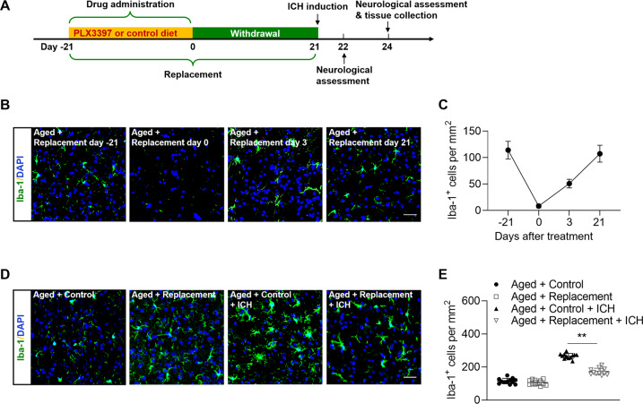
Fig. 1. Microglial replacement in the aged brain.
Groups of aged mice received 21 days of PLX3397 treatment in chow or a control diet, and then followed by three weeks of withdrawal prior to ICH induction. ICH induction, neurological assessment and tissue collection were conducted at indicated time points. A Flow chart illustrates drug administration and experimental design. B Brain sections from groups of aged mice receiving PLX3397 in chow at indicated time points after treatment or withdrawal were stained with Iba-1 (green). Nuclei were stained with DAPI (blue). Scale bar: 50 µm. C Counts of Iba-1+ cells in indicated groups of mice. n = 8 per group. D Brain sections from groups of aged mice receiving indicated treatments were stained with Iba-1 (green). Nuclei were stained with DAPI (blue). Scale bar: 50 µm. E Counts of Iba-1+ cells in indicated groups of mice. n = 12 per group. Data are presented as mean ± SD. **p < 0.01.

