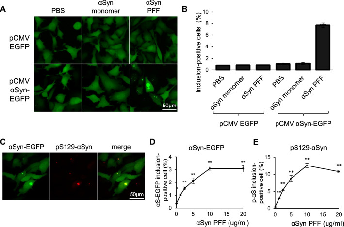
Figure 2.
Establishment of a cell-based assay to evaluate αSyn aggregation. (A) Representative images of αSyn-EGFP aggregation in HeLa cells. HeLa cells were transfected with the indicated plasmids, and then treated with PBS, αSyn monomers, or PFF. (B) Quantification of the percentage of cells containing obvious αSyn-EGFP inclusion bodies in (A). Data are shown as the mean ± SEM of twelve independent wells (n = 12; **P < 0.01; two-way ANOVA with the Tukey test). (C) Representative immunocytochemistry images of PFF-treated HeLa cells. Cells were transfected with pCMV αSyn-EGFP, and then treated with αSyn PFF. Phosphorylated αSyn (pS129 αS) was stained using a specific antibody. (D,E) Quantification of the percentage of cells containing obvious αSyn-EGFP (D) and p129-αSyn (E) inclusion bodies. Data are shown as the mean ± SEM of four independent wells (n = 4; *P < 0.05; **P < 0.01; one-way ANOVA with the Dunnett test compared with the no αSyn PFF control).

