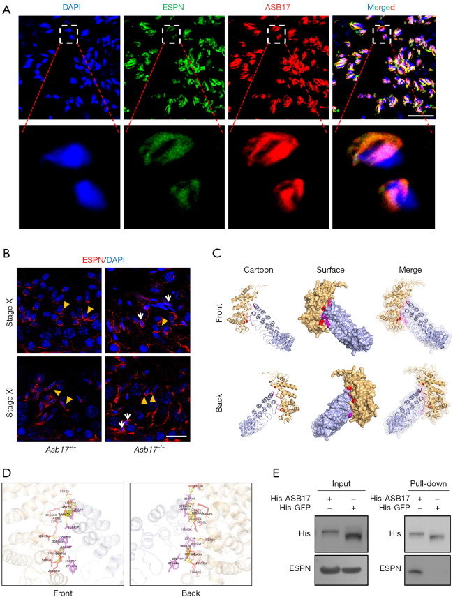
Figure 4.
ASB17 interacts with ESPN. (A) Co-immunostaining of ASB17 and ESPN in testes. Scale bar, 20 µm. (B) Immunostaining of ESPN in Asb17+/+ and Asb17−/− testes. Elongating spermatids are indicated by yellow arrowheads, while the retained mature spermatozoa in Asb17−/− testes are indicated with white arrows. Scale bar, 20 µm. (C) Predicted 3D structures of ASB17–ESPN complexes. The cartoon mode represents the backbone and secondary structures. The surface mode represents the solvent accessible surface area. ESPN and ASB17 are labeled as orange and blue, respectively. The binding residues between ESPN and ASB17 are labeled as red and purple, respectively. (D) Magnification of the binding interface. (E) Protein pull-down assay. His-tagged ASB17 and its negative control (his-tagged GFP) were purified from E. coli with a Ni-NTA Resin and incubated with testicular lysates. The protein complexes were resolved by SDS-PAGE and detected by western blot analysis. ASB17, Ankyrin repeat and SOCS box protein 17; ESPN: Espin.

