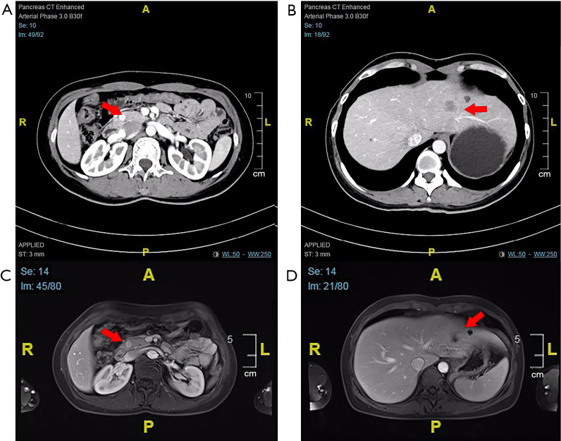
Figure 1.
CT scan and MRI. (A) The enhanced CT scan reveals a markedly enhanced lesion in the pancreatic head (1.0 cm ×0.9 cm). (B) A low-density lesion with slightly enhanced on the periphery of liver in the enhanced CT imaging. (C) MRI revealed a focal nodule with obvious enhancement in the pancreatic head. (D) A nodule (approximately 17 mm) with edge intensifying in arterial phase was found in in the left hepatic lobe on MRI. The arrows indicate the primary tumor in the head of pancreas and the metastatic liver lesion, respectively. CT, computed tomography; MRI, magnetic resonance imaging.

