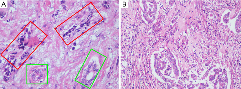
Figure 3.
Histopathological findings. (A) The tissue of pancreatic lesion stained with HE (field of view: 10×40). The neuroendocrine component consisting of neuroendocrine tumor and nonneuroendocrine part consisting of adenocarcinoma are in the red-framed and light green-framed areas, respectively. (B) The tissue of hepatic lesion stained with HE (field of view: 10×20). HE, hematoxylin and eosin.

