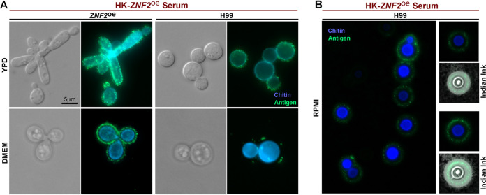
FIG 3.
The antigens are localized within the capsule and outside of the cell wall. (A) The ZNF2oe strain and the wild-type H99 were cultured in YPD medium at 30°C overnight (upper panel) or in DMEM medium at 37°C for 2 days in ambient air (lower panel). The cells were then used for immunofluorescence and probed with sera from mice vaccinated with HK-ZNF2oe cells (green). The cells were co-stained with calcofluor white to reveal chitin in the cell wall (blue). All images were taken at the same exposure. (B) The wild-type H99 cells were cultured in RPMI medium for 2 days. The cells were then used for immunofluorescence and labeled with sera from mice vaccinated with HK-ZNF2oe cells (green). The cells were co-stained with calcofluor white to reveal chitin in the cell wall (blue). Then the cells were negatively stained with Indian ink to reveal the capsule (white halo surrounding the yeast cells).

