
Figure 1. A schematic overview of the included P‐tau assays.
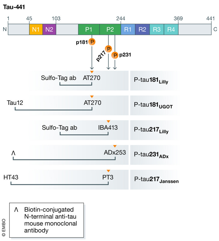
Schematic illustration of full‐length tau‐441, including N‐terminal, proline‐rich region, microtubuli binding domain, and C‐terminal. Anti‐tau antibodies are indicated for each of the five included P‐tau assays under the respective epitope region. P‐tau181UGOT is the P‐tau181 assay from the University of Gothenburg, as detailed in Karikari et al (2021).
For P‐tau231ADx, the inverted V symbol represents a biotin‐conjugated N‐terminal anti‐tau mouse monoclonal antibody, as detailed in Ashton et al (2021c).