
Figure 4. Expression of p27 on the surface of RSV‐infected A549 cells.
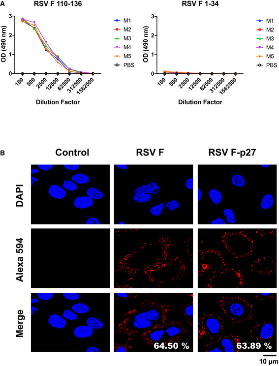
- Serum samples collected from individual mouse (M 1‐5) immunized with F 110‐136 peptide were tested for antibody binding against F‐p27 (110–136) peptide or F 1‐34 peptide (control) in ELISA.
- A549 cells were infected with RSV (MOI = 0.1) for 16 h, and fixed. Cells were treated with mock control rabbit sera (left panels), or rabbit antisera against F (center panels), or against F‐p27 (110–136) peptide (right panels), followed by Alexa 594 conjugated anti‐Rabbit IgG (red). Nuclei are stained with DAPI (blue). Scale bar = 10 µm. The number of cells positive for RSV‐F (middle panel) and RSV F‐p27 (right panel) upon counting of 200 cells were used to calculate percentage of cells stained for each antibody staining are shown in the ‘merge’ panel. The experiments were performed twice, and variation between the two independent experiments was < 6%.
Source data are available online for this figure.