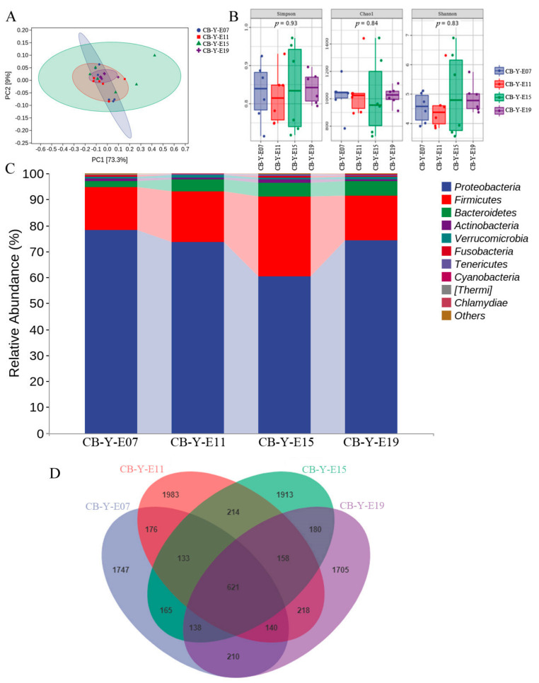
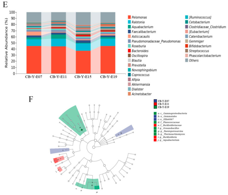
Figure 2.
Embryonic yolk microbial difference at different developmental stages. CB in the picture represents commercial broilers and Y represents yolk. (A) PCA: Each point represents a sample, and points of different colors indicate different groups. (B) Each panel corresponds to an alpha diversity index, which is identified in the gray area at the top. In each panel, the abscissa is the group label, and the ordinate is the value of the corresponding alpha diversity index. (C) The relative abundance of yolk microbiota in the chicken embryo at different days. (D) The Venn diagram shows the core microbes shared at different stages of development. (E) The relative abundance of yolk microbiota in the chicken embryo at different days (at the genus level). (F) Taxonomic cladogram generated from LEfSe showing significant difference in the microbiota profile of 4 stages of development.


