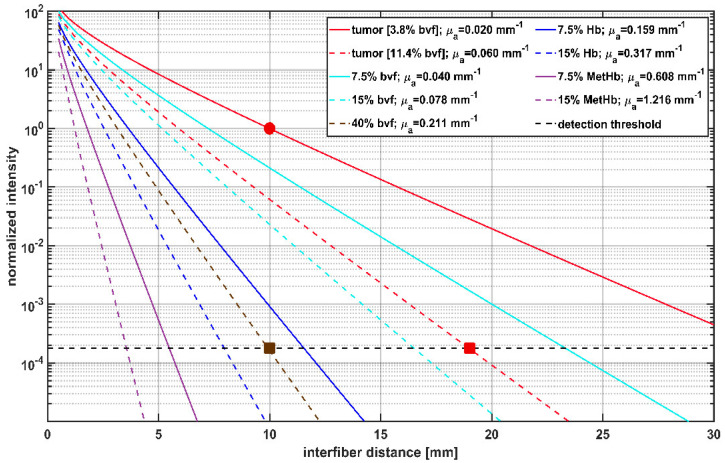
Figure 4.
Calculation of transmitted light intensity at 635 nm between cylindrical diffuser fibers (CDFs) with a 2 cm diffuser length, and its dependency on the interfiber distances for different blood volume fractions (bvf) and hemoglobin species. Blood is approximated to contain 85% HbO2 and 15% Hb. Transmitted light intensities are normalized to tumor (μa = 0.02 mm−1, μs’ = 2 mm−1) at 10 mm interfiber distance (red circle). The legend shows the different blood volume fractions of hemoglobin species and the resulting μa assumed for the calculation with μs’ set constant to 2 mm−1. The horizontal dashed line at 1.9 × 10⁻⁴ shows the estimated detection threshold of the iPDT SOM setup. The red square indicates that in case of a tumor with 11.4% bvf the detection threshold is reached at an interfiber distance of 19 mm. The brown square shows that for 40% bvf the detection threshold is already reached at an interfiber distance of 10 mm.

