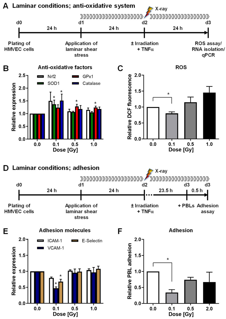
Figure 1.
Low-dose X-irradiation modulates mRNA expression of anti-oxidative factors, reduces reactive oxygen species (ROS) and peripheral blood lymphocyte (PBL) adhesion to human microvascular endothelial cells (HMVEC) under laminar conditions. (A) Experimental setup: For analysis of the mRNA expression of anti-oxidative factors, ROS and adhesion molecules under shear stress, laminar flow conditions were applied 24 h after plating of HMVEC. (B) After irradiation and stimulation with tumor necrosis factor alpha (TNF-α), RNA was isolated to measure the mRNA expression of anti-oxidative factors (Nrf2, Nuclear factor-erythroid-2-related factor 2; SOD1, Superoxide Dismutase 1; GPx1, Glutathione Peroxidase 1; Catalase) by quantitative PCR (qPCR) (mean + SEM; n = 6) or (C) cells were subjected to analysis of ROS by flow cytometry (mean + SEM; n = 8–12). (D) For adhesion assays, PBL were added 23.5 h after irradiation and adhesion was allowed for 0.5 h under constant laminar flow. (E) Measurement of adhesion molecules vascular cellular adhesion molecule (VCAM)-1, intercellular adhesion molecule (ICAM)-1, and E-selectin expression was performed by qPCR (mean + SEM; n = 6). (F) For adhesion assays, HMVEC were plated on glass cover slips, PBL were stained with CellTracker Green CMFDA (5-chloromethylfluorescein diacetate) and added 23.5 h after irradiation for 30 min to the medium reservoir under permanent laminar flow. HMVEC cells and adhered PBL were fixed and stained with 4′,6-diamidino-2-dhenylindol (DAPI) and phalloidin-tetramethylrhodamine B isothiocyanate (phalloidin-TRITC). PBL adhesion was evaluated by microscopic counting of PBL relative to the number of endothelial cells (mean + SEM; n = 5–9). * p < 0.05, Kruskal-Wallis test vs. 0 Gy.

