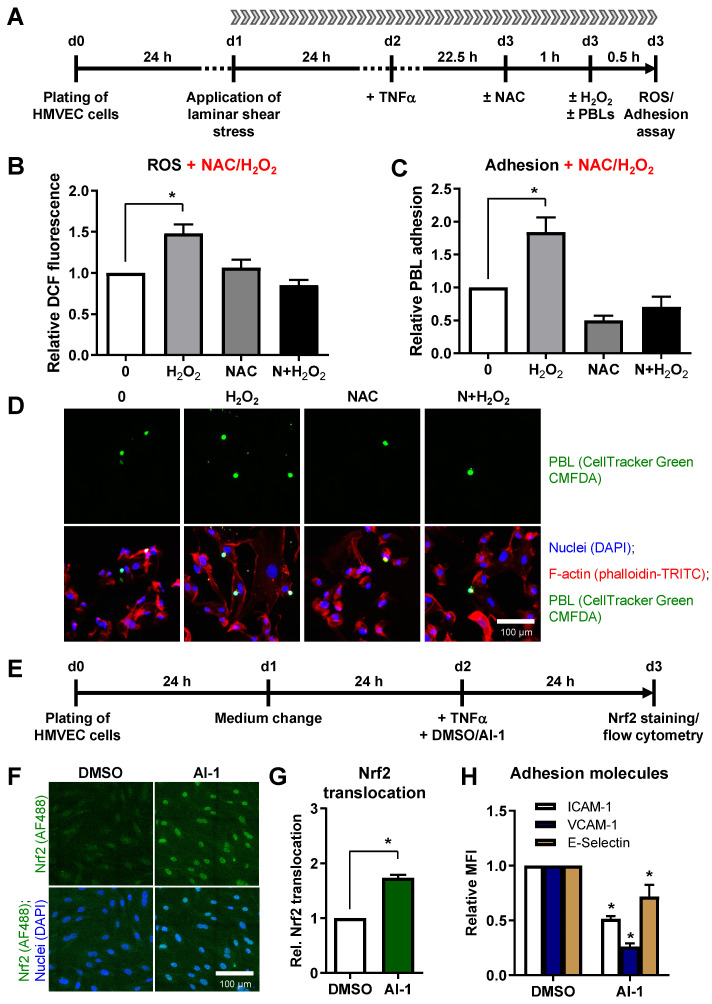
Figure 6.
Oxidative stress induces leukocyte adhesion to HMVEC under shear stress and Nrf2 activation decreases adhesion molecule surface expression. (A) Experimental scheme: HMVEC were plated and laminar shear stress was applied 24 h before TNF-α stimulation. After 22.5 h, cells were either treated with the ROS scavenger N-acetyl-L-cysteine (NAC) or left untreated. Oxidative stress was induced 1 h later by addition of hydrogen peroxide either without (H2O2) or with pretreatment with NAC (N+H2O2). (B) ROS measurement was subsequently acquired by flow cytometry (Mean + SEM; n = 6; * p < 0.05, one-way ANOVA vs. 0 Gy). (C) For adhesion experiments, Cell Tracker Green-stained PBL were added to the medium reservoir of the flow chamber and adhesion to HMVEC was allowed for 30 min under laminar flow (Mean + SEM; n = 6; * p < 0.05, one-way ANOVA vs. 0 Gy). (D) Exemplary pictures, depicting adhered PBL (CellTracker Green CMFDA (5-chloromethylfluorescein diacetate)), either alone or merged with nuclei (DAPI, blue) and F-actin staining of HMVEC cells (phalloidin-TRITC, red). Bar, 100 µm. (E) To analyze the impact of Nrf2 activation on adhesion molecule expression, HMVEC were plated, medium was changed after 24 h and cells were treated with TNF-α and DMSO or the Nrf2 activator AI-1. After 24 h, cells were fixed, stained for Nrf2 (AF488, green) and DAPI (blue). (F) Depicted are exemplary pictures, showing Nrf2 staining alone (green channel) or merged with nuclei (DAPI, blue). (G) Samples were evaluated microscopically for Nrf2 translocation, or (H) adhesion molecules ICAM-1, VCAM-1 and E-Selectin were measured by flow cytometry and values calculated as MFI + SEM relative to DMSO-treated cells. n = 4; * p < 0.05 (Mann-Whitney U test vs. DMSO).

