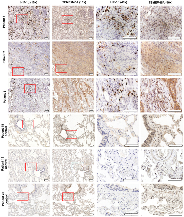
Figure 9.
The immunohistochemistry showed high expression of TMEM45A and sporadic expression of HIF-1α in invasive acinar lung adenocarcinoma. An area of higher magnification (40× objective) within the presented image of the region of interest at lower magnification (10× objective) is indicated by a red box. Stages were the following: for patient 1, T1bN2M0; patient 2, T3N0M0; patient 3, T2bN2M0. Control tissues were derived from pneumothorax nontumorous patients. Scale bar: 100 µm.

