Figure 5.
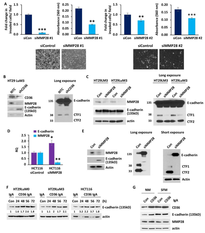
MMP28 reduces CRC cell invasion and decreases expression of functional E-cadherin in vitro. (A) Fold change in invaded cells/field, absorbance at 560 nm of the de-stained Matrigel trans-well invasion chambers and raw images of the Matrigel trans-well invasion chambers of the HCT116, siControl and siMMP28 cell lines (n = 3). (B) Western blot analysis of the HT29 LuM3 NTC and shCD36 cell lines for CD36, MMP28 and E-cadherin. The long exposure of the same Western blot for E-cadherin is also shown. (C) Western blot for MMP28 and E-cadherin expression in HT29LM3 and HT29LuM3 cells. Western blot including the E-cadherin cleavage products CTF1 and CTF2 is shown on a separate blot (long exposure). (D) qRT-PCR analysis of the control and MMP28 siRNA transfected HCT116 cell lines for MMP28 and E-cadherin. (E) Western blot for MMP28 and E-cadherin expression in HCT116 cells. Western blot including the E-cadherin cleavage products CTF1 and CTF2 is shown on separate blots (long and short exposure). (F) Western blot analysis showing the effect of CD36 blocking antibody on E-cadherin expression in HT29 LuM0, HT29LuM3 and HCT116 cells. Expression of E-cadherin is quantified based on band intensity and normalized to actin. (G) Western blot analysis showing the effect of CD36 blocking antibody on e-cadherin and MMP28 expression in HCT116 cells treated with IgA or IgCD39 for 48 h in normal medium or SFM (* p < 0.05, ** p < 0.01, *** p < 0.001).

