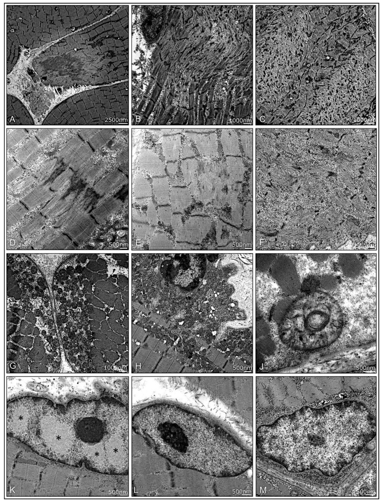
Figure 6.
Ultrastructural pathology in muscle biopsies from patients with jIIM. A high variability of disruption of myofibrillar architecture can be present in all subtypes. Focal, core-like alterations of the sarcomeric architecture in DM (P10) (A), P4 (B), and P12 (C). Z-band alterations with Z-band streaming in P15 (D) and dissolving Z-bands in anti-Jo1-ASyS (P14) (E) and focal glycogen deposits in OM (P7) (F). Moderate mitochondrial pathology with some mitochondrial subsarcolemmal aggregation in PL-7-ASyS (P9) (G) and DM (P2) (H). Few mitochondria with increased variability of diameter and altered cristae structure in P9 (J). Myonuclei with characteristic filamentous nuclear inclusions were only found in anti-Jo1-ASys (P14); note the different patterns of euchromatin, heterochromatin, and the nucleolus, as compared to the nuclear inclusions (*) (K). No inclusions were found in PL-7-ASyS (P6, P9) (L,M). (DM = dermatomyositis; ASyS = antisynthetase syndrome; OM = overlap myositis).

