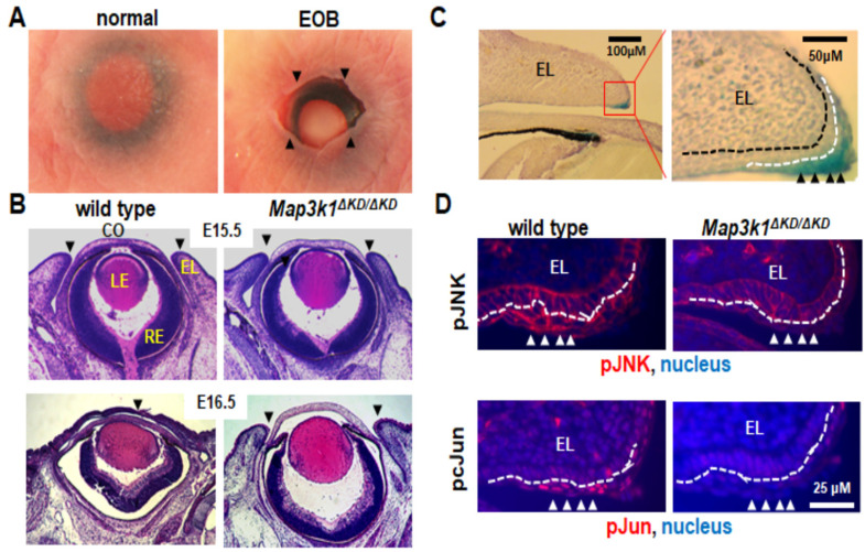
Figure 2.
Mouse embryonic eyelid development and closure. (A) Photographs of eyes of the newborn pups. Mice are normally born with eyelids closed, but those with defective embryonic eyelid closures are born with an EOB phenotype. Arrowheads point at the eyelid opening margins in EOB mice. (B) Histological analyses of the embryonic eyes at E15.5 and E16.5. At E15.5, the wild-type and Map3k1ΔKD/ΔKD embryos have the same eye structures, with the upper and lower eyelids separated. At E16.5, the eyelids were fused in the wild-type, but remained separated in the Map3k1ΔKD/ΔKD fetuses. Arrowheads point at the leading edge or fusion junction of the upper and lower eyelids. (C) Eye sections of the whole-mount X-gal stained Map3k1+/ΔKD E15.5 embryos were photographed at 10× (left panel), the red box in the left panel was shown at 40× magnifications (right panel). Abundant β-gal positive, i.e., MAP3K1-expressing cells were detected in the eyelid epithelium, particularly in the inferior periderm near the eyelid tip. (D) Immunohistochemistry analyses with antibodies for the pJNK (upper panels) and p-cJun (lower panels) of the E15.5 eyes. The pJNK and pJun were detected in the inferior periderm near the eyelid tip in wild-type but not Map3k1ΔKD/ΔKD embryos. Nuclei were stained with DAPI (blue). EL: eyelid, CO: cornea, LE: lens, RE: retina. Black dotted lines mark the basement membrane; white dotted lines mark the boundary between the basal epithelium and periderm. Arrowheads point at the MAP3K1-expressing periderm.

