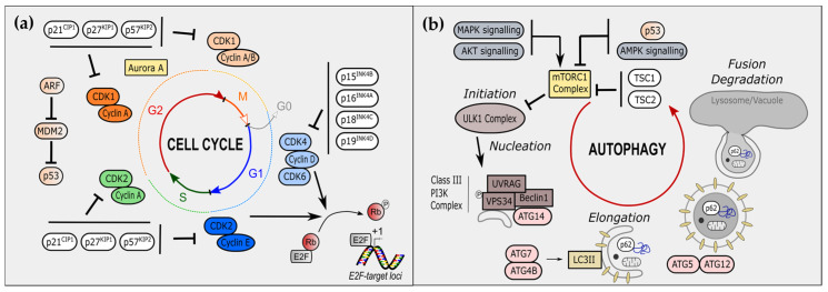
Figure 1.
Cell-cycle regulators and autophagy players. (a) Overview of the main cell-cycle regulators. The cell cycle is divided into four steps: G0 (grey)/G1 (blue), S (green), G2 (red), and M (orange) phases. Cyclin-dependent kinases (CDKs) associate with cyclins along the cell cycle, allowing its progression. In early G1, CDK4 and CDK6 bind to cyclin D. In late G1, CDK2 is associated with Cyclin E. CDK4/6-cyclin D and CDK2-cyclin E phosphorylate RB leading to E2F release and transcription at E2F-target loci of cell-cycle associated genes. Cyclin A associates with S-phase CDK2 and G2/M-phase CDK1. CDKs are inhibited upstream by cyclin-dependent kinase inhibitors (CKIs). In G1 phase, INK family including p15INK4B, p16INK4A, p18INK4C and p19INK4D, inhibits CDK4/6-cyclin D. In other phases, CIP/KIP family including p21CIP1, p27KIP1 and p57KIP2. Additional cell-cycle regulators include the Aurora Kinase A and the ARF/MDM2/p53 axis. (b) Overview of steps and actors of autophagy, called macro-autophagy. Initiation is mediated by the ULK1 complex, which mediates phosphorylation of VPS34 and formation of phagophore through nucleation and the association of UVRAG, Beclin-1 and ATG14. Elongation of phagophore is allowed by LC3II formation, by ATG7 and ATG4B, and LC3II association, by ATG5 and ATG12. Proteins can be also specifically targeted to phagophore by p62. Organelles, such as mitochondria, can be Fusion of phagophore with lysosome constitutes the last step, ultimately followed by degradation of internal components.

