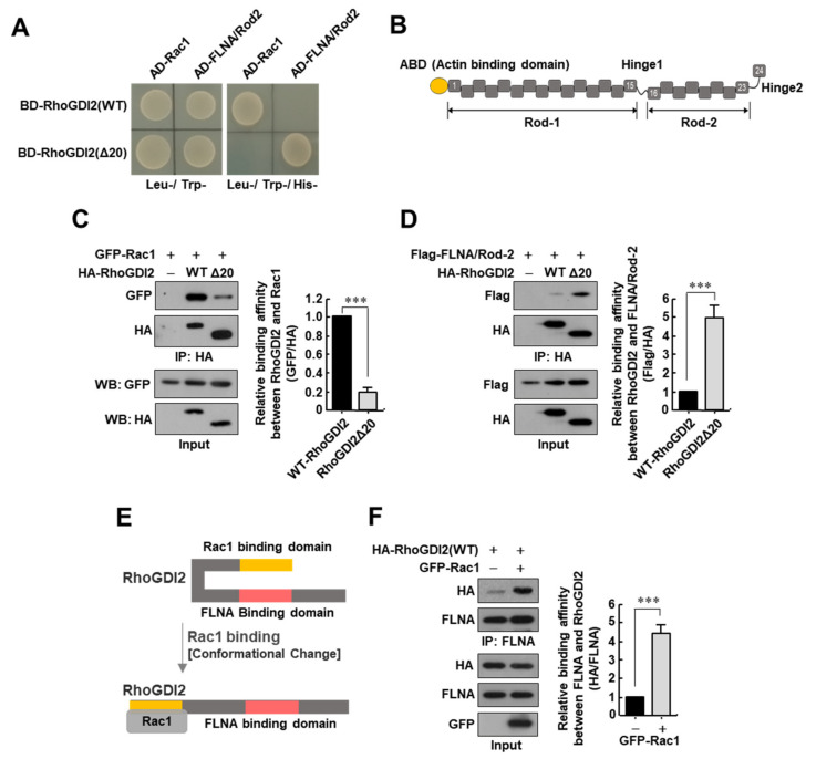
Figure 1.
Interaction between RhoGDI2 and Rac1 is a prerequisite for RhoGDI2 to interact with Filamin A. (A) Yeast two-hybrid experiments to see the interactions between wild type RhoGDI2 or RhoGDI2Δ20 and the Rac1 or Rod2 domain of Filamin A. (B) Schematic diagram of Filamin A structure. ABD; actin-binding domain, Rod-1 and Rod-2; immunoglobulin-like tandem repeat domains. (C) Interaction between exogenous Rac1 with wild type RhoGDI2 or RhoGDI2Δ20. HEK293T cells transfected with indicated constructs were immunoprecipitated and analyzed by Western blot (left). The data are representative of three independent experiments, and relative GFP and HA levels were quantified using ImageJ software (Version 1.53n, NIH, Bethesda, MD, USA) (right). ***, p < 0.001 as determined by t-test. The uncropped immunoblot images can be found in Figure S1. (D) Interaction between exogenous Rod2 domain of Filamin A (FLNA/Rod-2) and wild type RhoGDI2 or RhoGDI2Δ20. HEK293T cells transfected with indicated constructs, immunoprecipitated and analyzed by Western blot (left). The data are representative of three independent experiments and relative Flag and HA levels were quantified using ImageJ software (right). ***, p < 0.001 as determined by t-test. The uncropped immunoblot images can be found in Figure S1. (E) Schematic diagram of the conformational changes in RhoGDI2 after binding to Rac1. (F) Interaction between endogenous Filamin A with exogenous RhoGDI2 in the presence or absence of Rac1. HEK293T cells transfected with indicated constructs were immunoprecipitated and analyzed by Western blot (left). The data are representative of three independent experiments and relative FLNA and HA levels were quantified using ImageJ software (right). ***, p < 0.001 as determined by t-test. The uncropped immunoblot images can be found in Figure S1.

