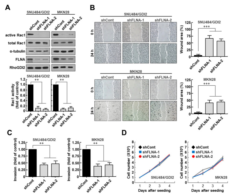
Figure 3.
Filamin A is required for Rac1 activation and invasive ability of gastric cancer cells. (A) Effect of Filamin A depletion on Rac1 activity in RhoGDI2-overexpressing SNU484 and MKN28 gastric cancer cell lines. Filamin A-depleted cell lysates were precipitated with a PAK1-agarose and analyzed by Western blot using anti-Rac1 antibody (upper). The data are representative of three independent experiments and ImageJ software was used to calculate the relative active Rac1 levels (lower). For normalization, total Rac1 and α-tubulin expressions were used as controls. **, p < 0.01 as determined by t-test. The uncropped immunoblot images can be found in Figure S5. (B) Effect of Filamin A depletion on migration ability of RhoGDI2-overexpressing SNU484 and MKN28 gastric cancer cells. A wound-healing assay was used to examine the migration ability of Filamin A-depleted SNU484/GDI2 and MKN28 cells, with wound closure visualized using phase-contrast microscopy (left). WimScratch software (Wimasis) was used to measure the wound areas. The percentage of the wound area is expressed as the means ± SD of three separate experiments (right). ***, p < 0.001 as determined by t-test. (C) Effect of Filamin A depletion on invasive ability of RhoGDI2-overexpressing SNU484 and MKN28 gastric cancer cell lines. Filamin A-depleted SNU484/GDI2 and MKN28 cells were cultured onto matrix-coated upper chambers and the number of invading cells was measured after 24 h. The data are presented as the means ± SD of three separate experiments, carried out in triplicate. **, p < 0.01 as determined by t-test. (D) Effect of Filamin A depletion on proliferation rate of RhoGDI2-overexpressing SNU484 and MKN28 gastric cancer cell lines. The indicated cells were cultured at a concentration of 1 × 10⁴ cells per well in a 6-well plate. After trypan blue staining, the viable cells were counted with a hemocytometer after 1 to 4 days of incubation.

