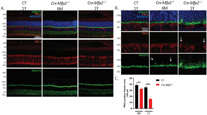
Figure 5.
Crx-Mfp2−/− mice show loss of bipolar cells and impaired synaptic connections. (A) PKCα (red) staining showed clear loss of bipolar cells at the age of 1 Y. (B) Higher magnification images revealed sprouting of bipolar cells (red) and VGLUT1 (green) mislocalization into the ONL. White arrows indicate the sprouting. (C) Quantification of PKCα positive bipolar cells per 100 µm revealed a significant loss in both 6-month-old and 1-year-old Crx-Mfp2−/− mice. N = 4/group. Statistical difference based on unpaired t-test: ** p < 0.01, **** p < 0.0001. Error bars indicate SD. RPE—retinal pigment epithelium; PR—photoreceptor; ONL—outer nuclear layer; OPL—outer plexiform layer; INL—inner nuclear layer; IPL—inner plexiform layer; GCL—ganglion cell layer.

