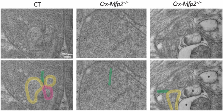
Figure 6.
Loss of photoreceptor ribbon synapse integrity in 6-month-old Crx-Mfp2−/− mice. Transmission electron microscopy images showed normal photoreceptor ribbon synapse formation in CT mice, with a ribbon (green), two horizontal cells (yellow) and a bipolar cell (pink). Crx-Mfp2−/− mice presented with free-floating photoreceptor ribbons and abnormal structures (*) in the photoreceptor synapses, which did not occur in CT mice. Top and bottom panel are the same images. Bottom panel includes interpretation. N = 4/group.

