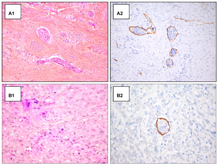
Figure A1.
H/E staining and D2-40 antibody staining in emboli detection. Examples of detection of lymphatic invasion in H&E staining and D2-40 antibody staining. (A) Detection of peripheric emboli in H&E staining (A1) and D2-40 antibody staining (A2) (original magnification 200×). (B) Unclear detection of intra-tumoral emboli in H&E staining (B1), after D2-40 antibody staining the intra-tumoral emboli detection appear clear (original magnification 400×) (B2).

