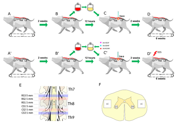
Figure 1.
Study design. Upper panel shows control animals: (A) Implantation of stimulating electrodes at Th5 and L2 followed by recovery and training of the mini-pigs (n = 4) to walk on a treadmill. (B) Collection of 50 mL of venous blood and preparation of the leucoconcentrate. (C) Spinal cord contusion injury followed by intravenous infusion of the autologous leucoconcentrate 4 h after surgery. (D) Training on a treadmill every second day starting 2 weeks after injury. Behavioural and electrophysiological data were collected over the next 6 weeks. Middle panel shows treated animals: (A’) Implantation of stimulating electrodes at Th5 and L2 followed by recovery and training of the mini-pigs (n = 4) to walk on a treadmill. (B’) Collection of 50 mL of venous blood and preparation of the genetically-enriched leucoconcentrate. (C’) Spinal cord contusion injury followed by intravenous infusion of the autologous genetically-enriched leucoconcentrate 4 h after surgery. (D’) Epidural electrical stimulation (EES) every second day combined with training on a treadmill starting 2 weeks after injury. Behavioural and electrophysiological data were collected over the next 6 weeks. Low panel: (E) The spinal cords were harvested at 8 weeks after injury. The rostral (RS) and caudal (CS) segments of the spinal cord relative to the epicentre of injury (red cross) were divided into three segments: RS1, RS2, RS3 and CS1, CS2, CS3, correspondingly, for histological and molecular studies. (F) Spinal cord areas used for histological investigation: ventral horn (VH) and lateral column (LC).

