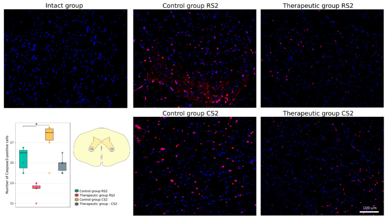
Figure 5.
Immunofluorescence staining of the ventral horns of the spinal cords in the rostral (RS2) and caudal (CS2) segments 8 weeks after contusion injury with an antibody against a pro-apoptotic protein (caspase-3) (red). Nuclei were counterstained with DAPI (blue). Morphometric analysis demonstrates the number of caspase3-positive nuclei in the intact, control and therapeutic groups in the corresponding areas, * p < 0.05. The squares inserted in the schematic transverse spinal cord image demonstrates the areas used for immunofluorescence analysis, VH—ventral horn.

