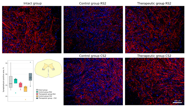
Figure 9.
Immunofluorescence staining of the ventral horns of the spinal cords in the rostral (RS2) and caudal (CS2) segments 8 weeks after contusion injury with an antibody against the synaptic vesicle protein synaptophysin (red). Nuclei were counterstained with DAPI (blue). Morphometric analysis demonstrates the mean (%) of the synaptophysin-positive area in the intact, control and therapeutic groups in the corresponding areas. The squares inserted in the schematic transverse spinal cord image demonstrates the areas used for immunofluorescence analysis, VH—ventral horn.

