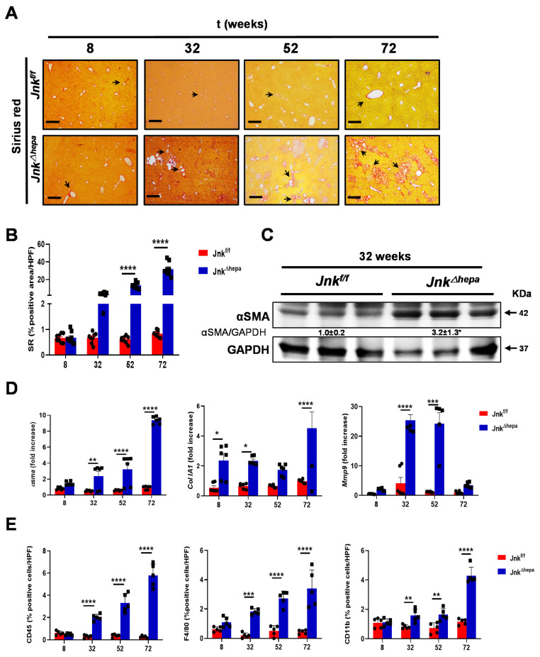
Figure 1.
Fibropolycystic disease in aging Jnk∆hepa mice is characterized by extracellular matrix deposition and inflammation. (A) Fibrosis was evaluated by SR staining in 8- to 72-week-old Jnkf/f and JnkΔhepa mice. Scale bars, 500 μm. (B) Quantification of SR areas was performed using ImageJ. Protein and mRNA expression was analyzed for αSma (left panel), ColIA1 (center panel) and Mmp9 (right panel) using Western Blot (C) and qRT-PCR (D), respectively (* p < 0.05; intergroup significance). (E) Quantification of positive cells from IF microphotographs of CD45 (left panel), F4/80 (center panel) and CD11b (right panel) is shown in 8- to 72-week-old Jnkf/f and JnkΔhepa mice. Data are shown as the mean ± SEM and graphed, separately (n = 6 mice per group) (* p < 0.05; ** p < 0.01; *** p < 0.001; **** p < 0.0001).

