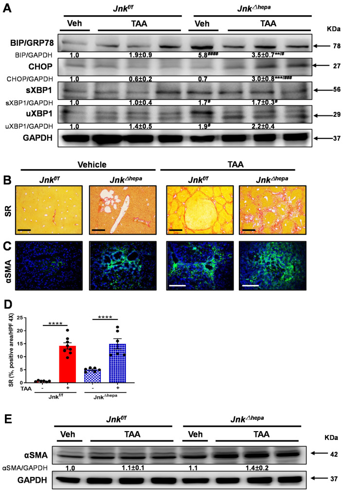
Figure 5.
TAA challenge exacerbates liver fibrogenesis in mice with hepatocytic deletion of Jnk1/2. (A) The expression of BiP/GRP78, CHOP, spliced XBP1 (sXBP1) and unspliced XBP1 (uXBP1) was evaluated in the livers of Jnkf/f and Jnk∆hepa mice treated or not with TAA using Western blot. Numbers denote molecular weight (KDa) of proteins. GAPDH served as loading control. (B) Representative Sirius Red (SR) stainings of liver paraffin sections from control and TAA-treated Jnkf/f and Jnk∆hepa mice (n = 6-8 mice per group). Scale bars, 500 μm. (C) Expression of αSMA protein was assessed via IF staining. Scale bars, 50 μm. (D) Positive area of fibrosis was calculated by ImageJ with microphotographs of SR staining. Data were represented as the mean ± SEM and graphed. (E) Expression of αSMA was analyzed by Western blot in the indicated groups of mice. Numbers denote molecular weight (KDa) of proteins. GAPDH served as loading control. (# p < 0.05; ** p < 0.01; ***/### p < 0.001; ****/#### p < 0.0001).

