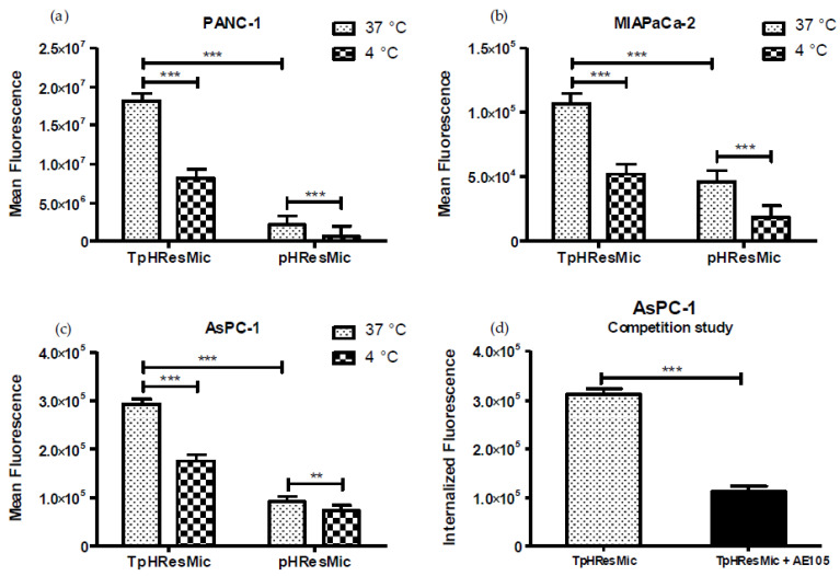
Figure 8.
(a–c) Uptake studies of the targeted (TpHResMic) and no targeted micelles (pHResMic) on 2D model of PDAC cells. The histogram plots report the mean fluorescence values obtained after incubation for 1 h of PDAC cells with BDP@TpHResMic and BDP@pHResMic at pH 6.8 medium and at 37 °C or 4 °C, and refer to three independent experiments. (d) The histogram plots report the mean ± SD of the fluorescence values obtained in the competition study of the targeted BDP@TpHResMic, with AE105 peptide in AsPC−1 cell line. (** p < 0.01, *** p < 0.001).

