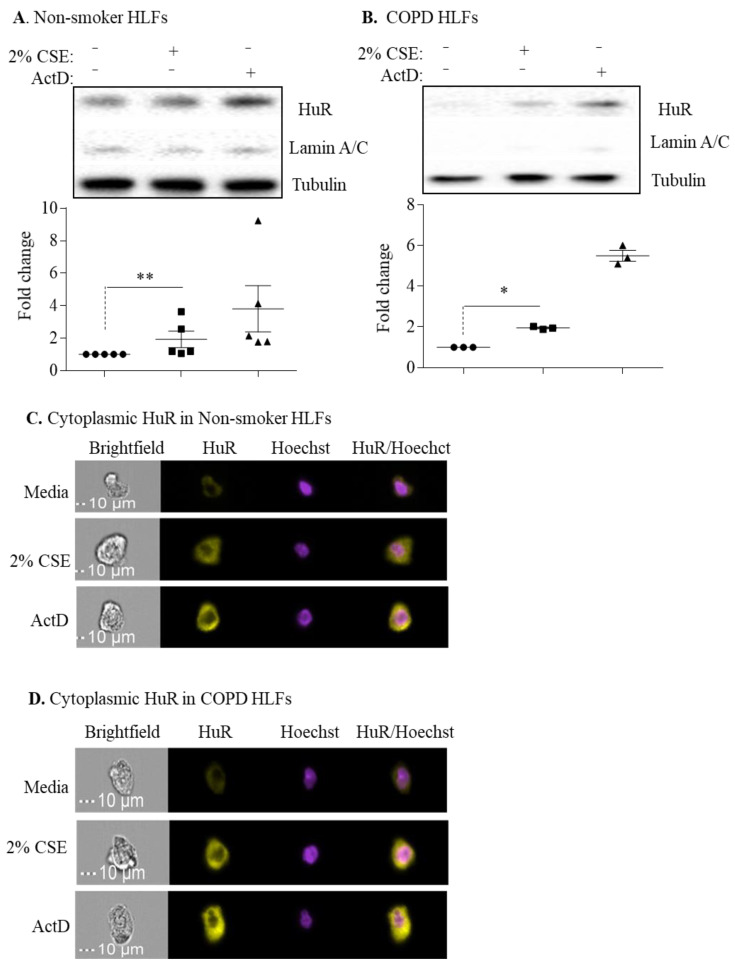
Figure 8.
Cytoplasmic HuR is increased by cigarette smoke. (A) Cytoplasmic HuR in non-smoker HLFs: there was an increase in HuR cytoplasmic localization in response to 2% CSE for 4 h (** p = 0.004). Actinomycin D (ActD) was used as positive control for the translocation of HuR. Lamin A/C is a nuclear marker, while tubulin is a cytoplasmic marker. Results are expressed as the mean ± SEM of 5 independent experiments. (B) COPD HLFs: there was an increase in HuR cytoplasmic localization in response to 2% CSE for 4 h (* p < 0.05). Results are expressed as the mean ± SEM of 3 independent experiments. Data between untreated and CSE-exposed cells were analyzed by a Mann–Whitney one-tailed t-test. (C) Cytoplasmic HuR in non-smoker HLFs: HuR localization in non-smoker HLFs treated with 2% CSE was assessed by Imaging Flow Cytometry. There was an increase in HuR expression in response to 2% CSE for 4 h. ActD was used as a positive control for HuR translocation into the cytoplasm. A representative picture for cells is shown from 2 independent experiments. (D) Cytoplasmic HuR in COPD HLFs: there was an increase in HuR expression in the cytoplasm of COPD HLFs exposed to 2% CSE for 4 h. A representative picture for cells is shown from one COPD subject.

