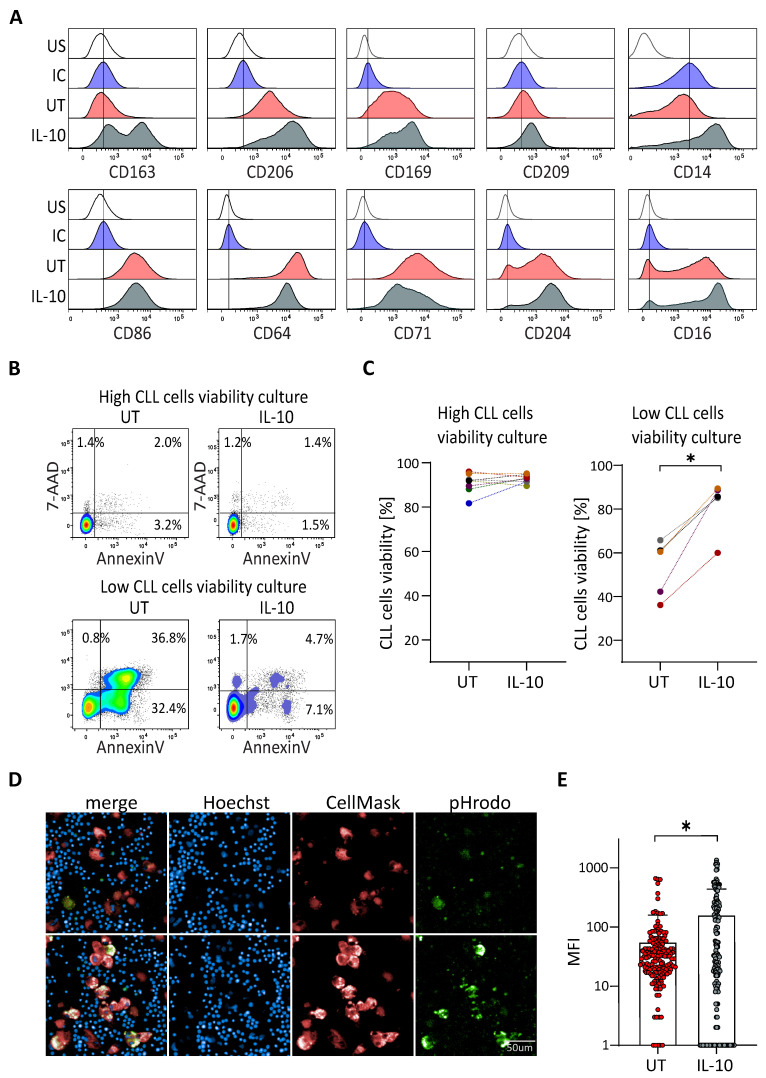
Figure 2.
IL-10 rescues viability of CLL cells from patients with low protective NLC. (A) Surface markers expressed by NLC analysed by flow cytometry at 14 days of cultures of PBMC from CLL patient with low in vitro CLL cells viability, untreated (UT: red) or treated with IL-10 (grey), comparing to the unstained (US: white) and the isotypic (IC: blue) controls. Representative histograms from five independent experiments. (B,C) Percentage of the CLL cells viability at 14 days of culture of PBMC from CLL patient with low or high in vitro CLL cells viability untreated (UT) or treated with IL-10 (B) one representative experiment; (C) eight and five independent experiments for high viability and low viability cultures respectively). (D,E) Phagocytosis of CLL cells (blue dots) by NLC (red) visualized (D) upper: untreated; lower: IL-10 treated and quantified (E) thanks to the green pHrodo fluorescence. * indicates p < 0.05.

