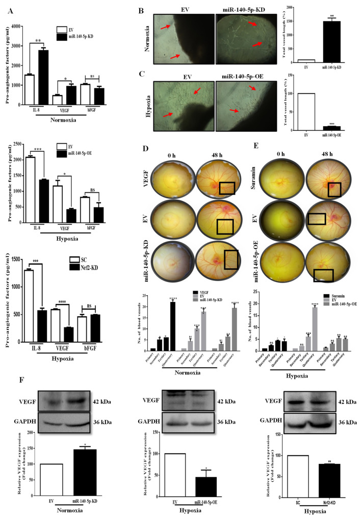
Figure 5.
miR-140-5p suppresses angiogenesis in BC. (A) ELISA was performed to measure secretion of pro-angiogenic factors (IL-8, VEGF, and bFGF) in CM from miR-140-5p-KD cells under normoxia, miR-140-5p-OE cells or Nrf2-KD cells under hypoxia. (B,C) Rat aortic ring sections cultured on matrigel containing CM from miR-140-5p-KD cells under normoxia or miR-140-5p-OE cells under hypoxia. The vascularized area is indicated by red arrowheads. Quantification of the vascular area was performed using AngioTool. (D,E) Representative images of the chick YSM assay treated with CM from either miR-140-5p-KD cells under normoxia or miR-140-5p-OE cells under hypoxia. Here VEGF and Suramin were used as positive and negative controls respectively to test the angiogenic potential of miR-140-5p. (F) Analysis of VEGF expression by Western blotting in miR-140-5p-KD cells under normoxia and miR-140-5p-OE cells or Nrf2-KD cells under hypoxia. Error bars indicate mean ± SEM (n = 3). Student’s t-tests were used to compare the means of two groups. ns: not significant, * p < 0.05, ** p < 0.01, *** p < 0.001, and **** p < 0.0001 compared to EV.

