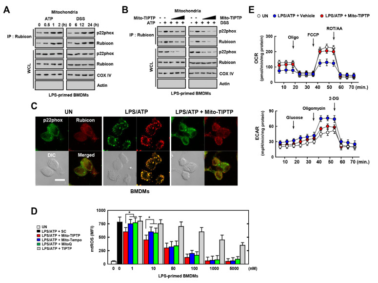
Figure 5.
Effects of Mito−TIPTP in mitochondrial functions. BMDMs were mitochondria fractionated by Mitochondria Isolation Kit. (A) LPS (100 ng/mL)-primed mitochondria fractionated−BMDMs were activated with ATP (1 mM) for 30 min or DSS (3%) for 18 h, and subjected to IP with αRubicon. IB with αp22phox and αRubicon. WCLs were used for IB with αRubicon, αp22phox, αCOX IV, and αActin. (B) The experimental conditions follow the same pattern as outlined in (A). LPS−primed BMDMs were treated with Mito−TIPTP (1, 10, and 100 nM) for 1 h, and then activated with ATP for 30 min or DSS for 18 h. (C) Immunostaining of BMDMs treated with LPS/ATP in presence or absence of Mito−TIPTP (10 nM) and then immunolabeled with antibody to p22phox (Alexa Fluor 488) or Rubicon (Alexa Fluor 568). Scale bar, 10 μm. (D) FACS analysis for mitoROS of BMDMs treated with LPS/ATP in the presence or absence of Mito−TIPTP, Mito−Tempo, or MitoQ in indicated concentrations. Quantitative analysis of mean fluorescence intensities of ROS (box). (E) The real−time measurement of OCR, as an indicator of oxidative metabolism (left) or ECAR, as an indicator of glucose oxidation. Data shown are representative of seven independent experiments with similar results (A–E). Statistical significance was determined by Student’s t−test with Bonferroni adjustment (* p < 0.05) compared with solvent control (0.1% DMSO).

