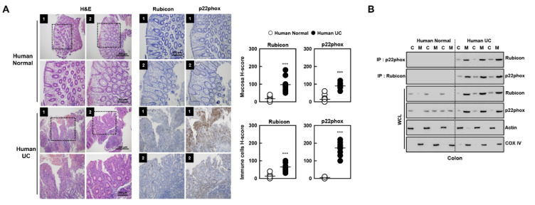
Figure 8.
H&E staining and immunohistochemistry in colon in human normal and ulcerative colitis (UC) patients. (A) Human normal and UC patients were used for H&E staining and IHC with αp22phox and αRubicon (left). H-score Rubicon and p22phox in mucosa and immune cells in colon were calculated by multiplying the percentage of the stained area by the staining intensity (right). Representative images from five independent healthy controls and patients are shown. Insets, enlargement of outlined areas. Biological replicates (n = 10) for each condition were performed. (B) Colon of human normal and UC patients were cytoplasmic and mitochondria fractions separated and analyzed for Rubicon, subjected to IP with αRubicon and αRubicon. IB with αp22phox and αRubicon. WCLs were used for IB with αRubicon, αp22phox, αCOX IV, and αActin. Data from three of ten normal human and UC patients are shown. Statistical significance was determined by Student’s t-test with Bonferroni adjustment (*** p < 0.001) compared with human normal.

