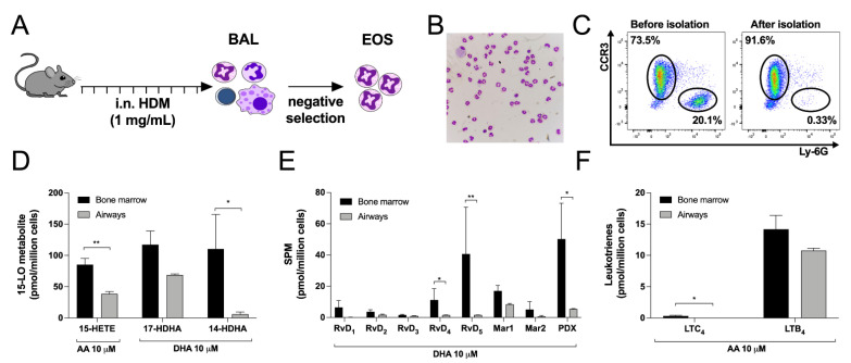
Figure 7.
Comparison between eosinophils isolated from the airways of HDM-treated mice and eosinophils derived from the bone marrow. (A) Experimental design to isolate mouse eosinophils from the airways of HDM-treated mice. (B) Morphology of airway cells after eosinophils enrichment determined by DiffQuik staining. (C) Purity of airway EOS before and after negative selection. (D–F) Eosinophils (5 × 10⁶ cells/mL, 37 °C) were treated with (D) 10 µM AA or 10 µM DHA and (E) 10 µM DHA or (F) 10 µM AA for 15 min. Incubations were stopped by adding one volume of cold (−20 °C) MeOH containing the internal standards. Samples were processed and analyzed for eicosanoids and docosanoids content by LC-MS/MS, as described in Materials and Methods. Results are the mean (±SEM) of 7 independent experiments using bone marrow-derived mouse eosinophils and 4 independent experiments using mouse eosinophils isolated from the airways. Data from bone marrow derived eosinophils are the same as those presented in Figure 1, Figure 3 and Figure 5. p-values were obtained as described in Materials and Methods. * p < 0.05, ** p < 0.01.

