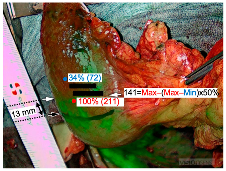
Figure 2.
Fluorescence imaging (FI) with indocyanine green (ICG). The maximum ICG signal was 211 (red marker) and the minimum was 72 (blue marker). The subjective transection line was drawn in black intraoperatively. The ICG-based resection line was obtained 13 mm peripheral to this line at a 50% decrease of the maximum ICG signal (white arrows).

