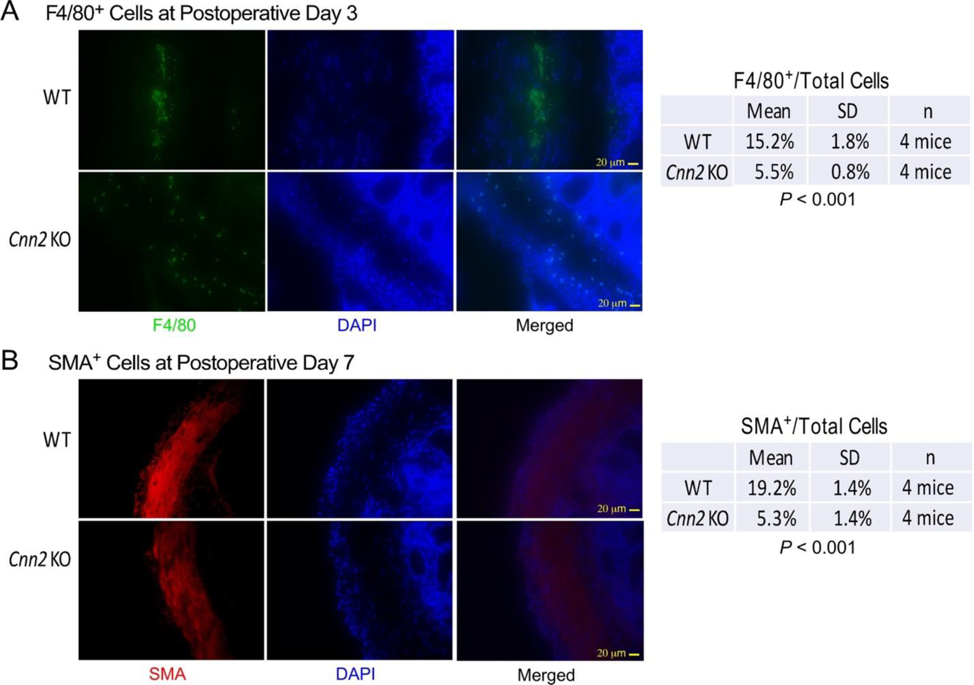
Figure 5: Calponin 2 KO mice exhibit attenuated macrophage infiltration and myofibroblast differentiation during the healing of operational injuries.

(A) Immunofluorescence micrographs of thin sections of cecal adhesion sites at postoperative Day 3 showed significantly less infiltration of F4/80+ macrophages (green) in calponin 2 KO mice in comparison with that of wild type controls. (B) At postoperative Day 7, immunofluorescence microscopy further showed significantly less α-SMA-positive (red) myofibroblasts in sections of cecal adhesion sites from calponin 2 KO mice than that in WT control. DAPI nucleus stain (blue) was used to identify the total number of cells.
