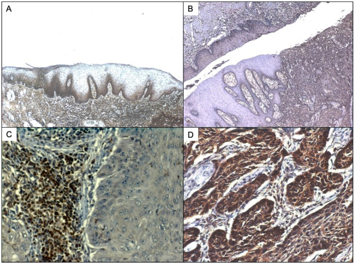
Figure 2.
(A) SP immunostaining of oral non neoplastic epithelium. SP can be detected in this case both in the membrane of basal keratinocytes and also in the nuclei of subepithelial lymphocytes. The stroma also presents positive inflammatory infiltrate. (B) NK1R immunostaining of laryngeal carcinoma and dysplastic epithelium. Strong immunostaining for NK1R can also be detected in the lymphocytes under the basal lawyer of mucosa and tumour infiltrating lymphocytes. (C). SP immunostaining of laryngeal carcinoma and dysplastic epithelium with greater increase to show better the infiltrating lymphocytes of dysplastic epithelia. (D) SP immunostaining of laryngeal carcinoma showing, under greater zoom, the infiltrating nests expressing SP.

