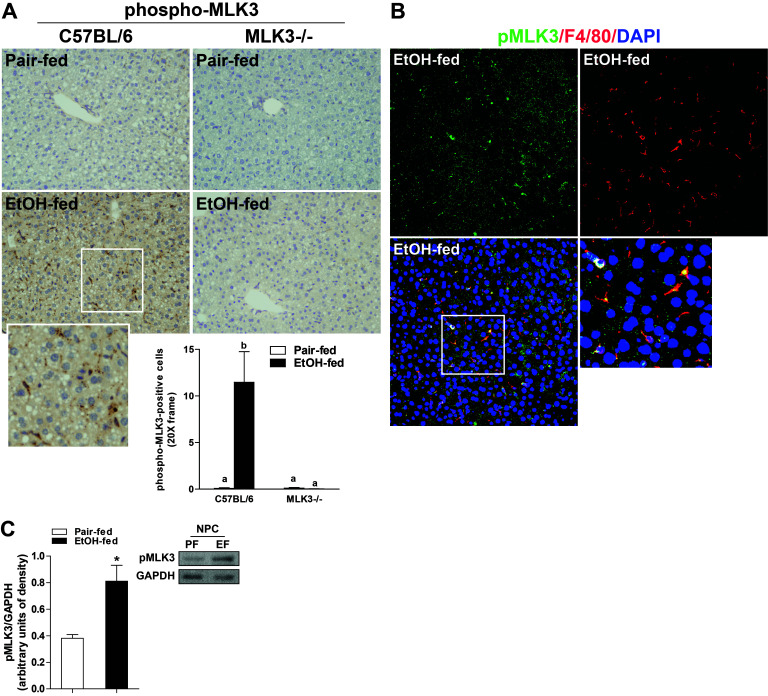
Figure 2.
Chronic ethanol feeding increased phosphorylation of MLK3 in mouse liver. C57BL/6 and MLK3−/− mice were allowed free access to diets with increasing concentrations of ethanol (final concentration 32% of kcal) or pair fed a control diet for 25 days. Paraffin-embedded livers were deparaffinized followed by immunohistochemistry. (A) Immunoreactive phospho-MLK3 was visualized, and nuclei were counterstained with hematoxylin. Images were acquired using 20× objective. Positive staining was quantified using Image-Pro Plus software and analyzed. Values represent means ± SEM, n = 4 pair-fed and 6 EtOH-fed mice. Values with different lower case letters are significantly different from each other, p < 0.05. (B) Immunoreactive phospho-MLK3 (green) and F4/80 (red) were visualized, and nuclei were labeled with DAPI. Images were acquired at 40×. (C) Nonparenchymal cells were isolated from livers of wild-type mice. Lysates were prepared and used for Western blot analysis of phospho-MLK3. GAPDH was used as a loading control. Values represent means ± SEM, n = 6–7 mice. *p < 0.05 compared to pair fed.

