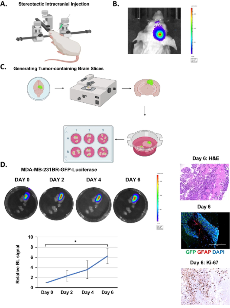
Figure 1. Intracranial injection, brain slice generation, and BCBM growth ex vivo.
(A) Schematic representation of mouse during intracranial injection under a stereotactic instrument. (B) Representative images of bioluminescent detection of tumors from 4–6 weeks old Nu/Nu mice injected with 5 × 10⁵ MDA-MB-231BR-GFP-Luciferase cells 12 days post-injection. (C) Schematic representation of the generation of ex vivo mouse brain slices from (B). (D) Representative images depicting tumor growth in organotypic brain slices derived from mice intracranially injected with MDA-MB-231BR-GFP-Luciferase cells detected via bioluminescence over 6 days. H&E & Ki-67, GFP (tumor cells) GFAP (reactive astrocytes) DAPI (nucleus) staining of a representative brain slice with tumor (image magnification 20x, scale bar: 200 μm). Quantification of tumor growth represents bioluminescence signal relative to day 0 (n = 3). Student’s t-test reported as mean ± SEM, * p < 0.05. Please click here to view a larger version of this figure.

