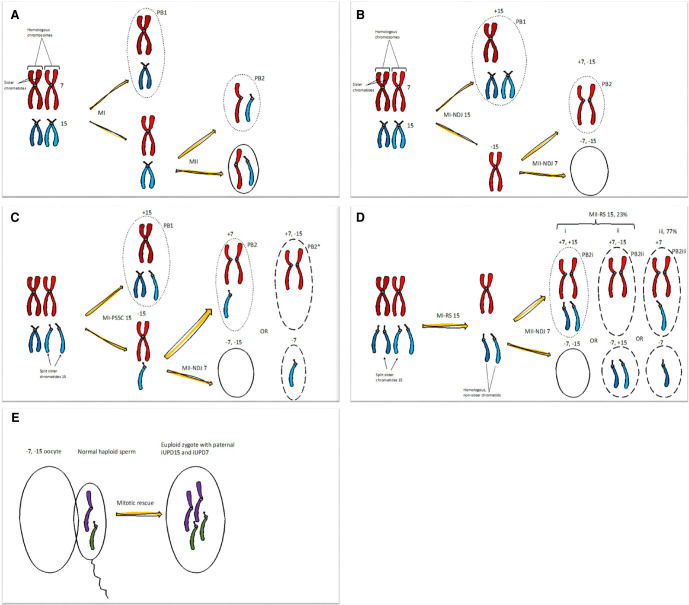
Figure 2.
Abnormal female meiosis resulting in a double nullisomic oocyte, in which alternative C or D is the most likely event in our case. (A) Normal situation with canonical meiotic division I and II with normal segregation of Chromosomes 7 (red) and 15 (blue). The first meiotic division separates the pair of homologous chromosomes, whereas the second division separates sister chromatids. Dotted lines represent polar bodies (PB), and complete lines the oocyte. Recombinations are omitted from the figures for simplicity. (MI) Meiosis I, (MII) meiosis II. (B) Canonical MI and MII with nondisjunction (NDJ). At MI, homologous chromosomes should segregate to opposite spindle pools, but here the homologous Chromosome 15 missegregate. Chromosome 15 is the most frequent chromosome involved in aneuploidy and also represents the chromosomes with the strongest maternal age effect on premature separation (and missegregation) of sister chromatids (PSSC) and reverse segregation (RS) (McCoy et al. 2015; Capalbo et al. 2017; Gruhn et al. 2019). In an aneuploid oocyte, the risk of MII-NDJ increases, here depicted with NDJ of Chromosome 7, where the sister chromatids fail to separate. The double nullisomy oocyte (−7, −15) outcome is outlined. Polar body 1 (PB1) from MI-NDJ show +15 (disomy 15), and PB2 (dotted line) show +7, −15 (mixed disomy and nullisomy). The result from MII-NDJ of Chromosome 7 in an oocyte with a PB1 constitution is not drawn but would be +7,+15 (double disomy) and −7, +15 (mixed nullisomy and disomy). (C) Meiosis with premature (or precocious) separation of sister chromatids (PSSC), in which sister chromatids of one Chromosome 15 loose cohesins and split prematurely and separate in MI, forming a free chromatid, and segregate with (PB1, +15) or without (−15) the homologous chromosome. In addition, we include MII-NDJ of Chromosome 7. Chromatid 15 can be expelled into PB2 (+7) in MII, making a nullisomic oocyte (−7, −15). This chromatid could also stay in the oocyte during MII; see the dashed outlines of the alternative oocyte (−7) and PB2* (+7, −15). (D) Noncanonical meiosis with reverse segregation (RS), in which sister chromatids of Chromosome 15 segregate to different primary oocytes in MI, and homologous chromatids segregate in MII (iii). For simplicity, we have drawn only one of the two outcomes and also omitted PB1. RS of Chromosome 15 occurs in MI, and the three possible MII outcomes depicted all show MII-NDJ of Chromosome 7. An additional RS-MII error of the non-sister chromatids 15 occurs in the first two alternatives (i and ii), and balanced segregation of 15 in the last (iii). According to Ottolini et al. (2015) MI-RS error with missegregation of the non-sister chromatids into the same oocyte in MII (i and ii) will occur in 23%. We have outlined the double nullisomic oocyte; the corresponding PB2 is marked with dots (i), and alternative oocytes and PB2s (ii, iii) with dashes. (E) Fertilization with a balanced haploid sperm, followed by postzygotic double monosomy rescue via endoduplication in the zygote, producing a double paternal isodisomy of Chromosomes 7 and 15. Maternal MII completion, including extrusion of PB2, occurs after fertilization with the haploid sperm, but this is drawn separately for simplicity.

